
The Unseen Masterpieces: Evident Scientific Unveils Winners of the 6th Annual Image of the Year Award

The intersection of rigorous scientific inquiry and breathtaking aesthetic beauty has once again been illuminated as Evident Scientific announced the winners of its 6th Annual Image of the Year Award. This prestigious competition, which celebrates the pinnacle of scientific light microscopy, serves as a global stage for researchers, clinicians, and enthusiasts to showcase the hidden wonders of the microscopic world. By transforming complex biological and material data into visual masterpieces, the award highlights the indispensable role of imaging technology in modern science.
This year’s winning entries span a diverse array of subjects—from the self-organizing complexity of human brain cells to the structural elegance of lignin fibers—proving that the "unseen" world is as vibrant and intricate as anything visible to the naked eye.
Main Facts: The Global and Regional Champions
The 6th iteration of the contest saw a record-breaking level of participation, with submissions hailing from 34 countries. Each entry was scrutinized by an international panel of experts who evaluated the images based on three primary criteria: artistic and visual aspects, scientific relevance, and microscopic proficiency.
The Global Winner: Mapping the Human Brain’s Architecture
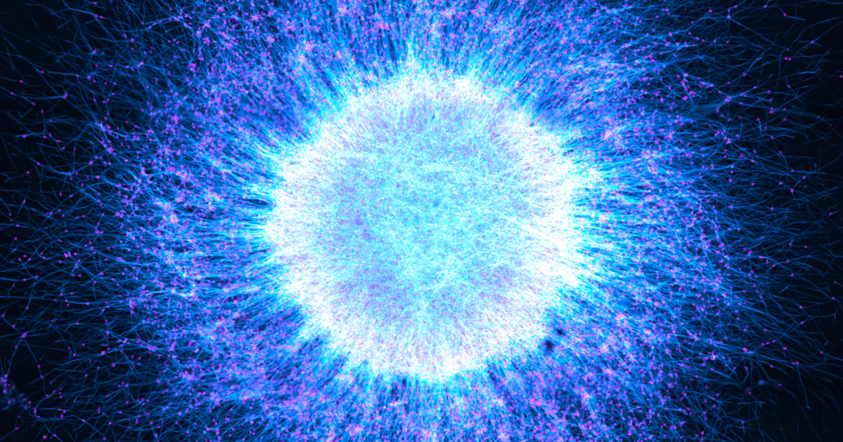
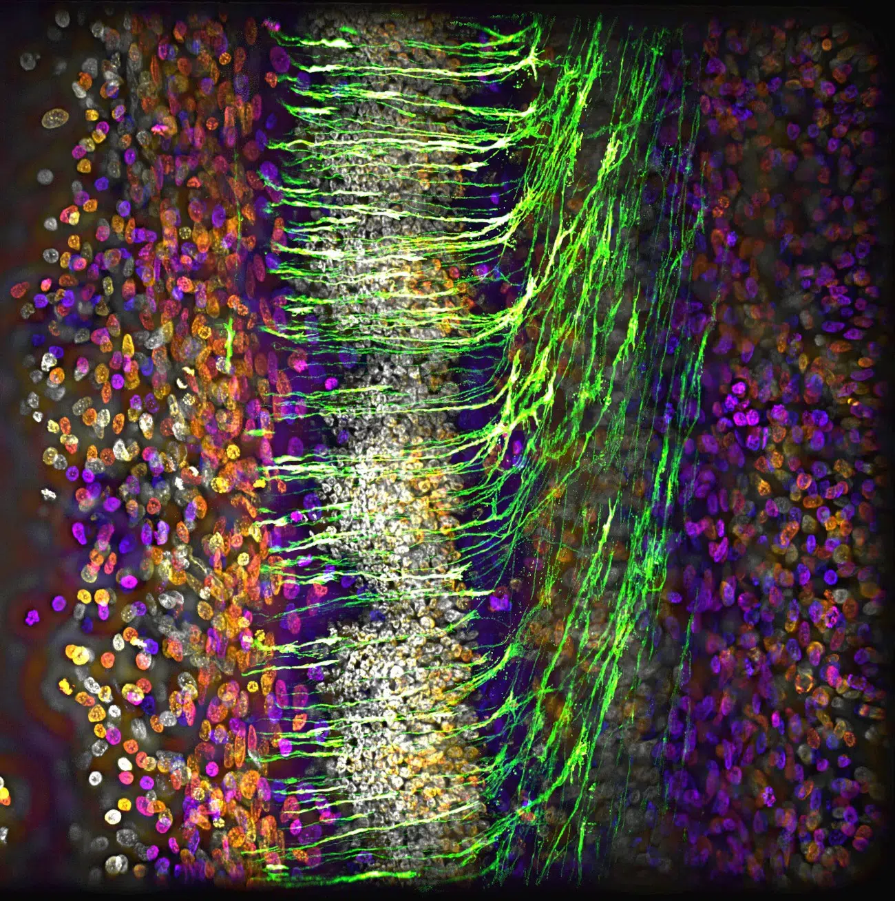
The top honor of Global Winner was awarded to Katie Holden of the United Kingdom. Her mesmerizing image features induced pluripotent stem cell (iPSC)-derived neurospheres. These are clusters of neural stem cells that have been reprogrammed from adult cells, offering a window into how the human brain develops.
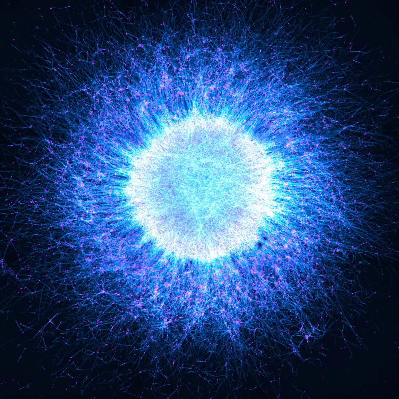
The image is more than just a vibrant display of colors; it represents a breakthrough in regenerative medicine and developmental biology. The neurospheres in Holden’s capture demonstrate how neuronal cells self-organize into structures that mirror the layered complexity of the human brain. This "mini-brain" model allows scientists to study neurological disorders and brain development without the need for invasive procedures. For her achievement, Holden was awarded a high-end Evident SZX7 stereo microscope paired with a DP23 digital camera, tools that are themselves industry standards for high-resolution imaging.
Materials Science: The Art of Industrial Anatomy
In the Materials Science category, Muhammad Tahir Khan of Ireland took the top prize for his striking image of lignin fiber. To the untrained eye, the image appears to be an aerial photograph of vast, wind-swept sand dunes bathed in a palette of orange and purple. In reality, it is a close-up of the organic polymer that provides structural support to the tissues of vascular plants and some algae. Lignin is crucial in the paper and bio-fuel industries, and Khan’s image highlights the structural integrity and hidden beauty of industrial materials.

Regional Excellence Across the Continents
The competition also recognizes regional leaders who pushed the boundaries of microscopy in their respective parts of the world:
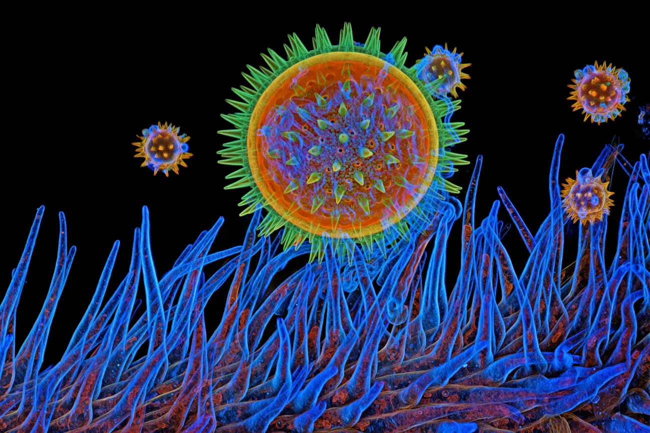
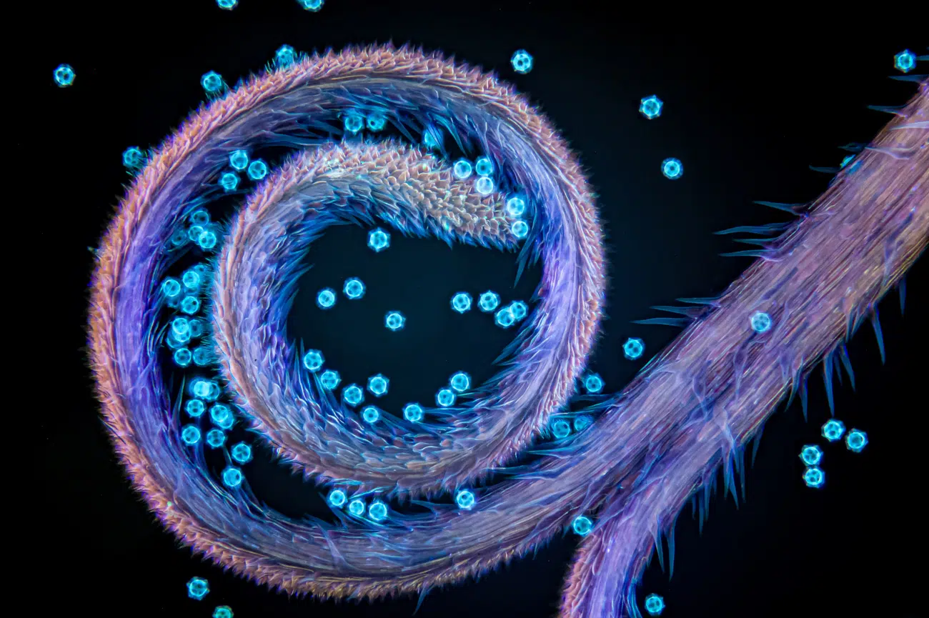
- Americas: Igor Siwanowicz (USA) won for his detailed capture of Mallow pollen resting on a stigma. Siwanowicz is a perennial favorite in microscopy circles, known for his ability to bring out the architectural majesty of botanical and insect life.
- EMEA (Europe, Middle East, and Africa): Gerd Günther (Germany) secured the award with an image of the stigma of a chicory plant, complete with pollen grains. The image emphasizes the delicate "lock and key" mechanisms of plant reproduction.
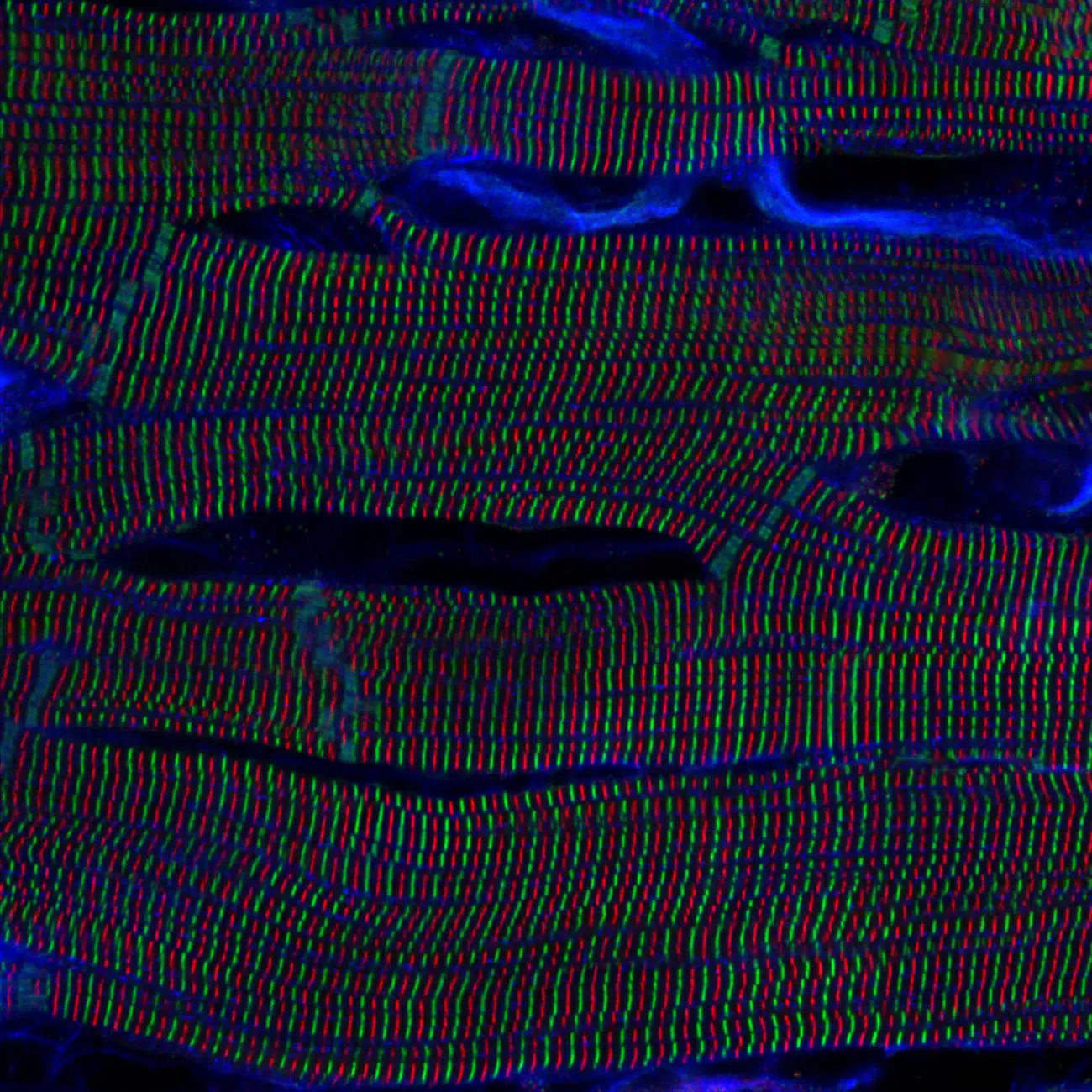
- Asia-Pacific: Kentaro Mochizuki (Japan) captivated the judges with a hypnotic, high-contrast shot of sarcomere structures within cardiomyocytes (heart muscle cells) of a rat heart. This image provides a clear view of the microscopic units responsible for every heartbeat.
Chronology: The Evolution of the Image of the Year Award
The Image of the Year Award began as the "Olympus Image of the Year Life Science European Award" before expanding into a global competition. Over the last six years, the contest has mirrored the rapid technological shifts in the microscopy industry.
- The Early Years (2018-2019): Initially focused on life sciences, the competition sought to bridge the gap between laboratory research and public engagement.
- Global Expansion (2020): The competition was restructured to include regional categories for the Americas, EMEA, and Asia-Pacific, reflecting the global nature of scientific collaboration.
- The Transition to Evident (2022-2023): Following the divestment of Olympus Corporation’s Scientific Solutions Division into the independent entity known as Evident Scientific, the award was rebranded. This transition allowed for a broader focus, leading to the inclusion of a dedicated Materials Science category to recognize the importance of microscopy in engineering and chemistry.
- The 6th Annual Milestone (Current): This year’s contest marks a peak in technical sophistication. The use of advanced staining techniques, confocal microscopy, and super-resolution imaging has allowed participants to capture biological processes with unprecedented clarity.
Supporting Data: Technical Precision and Global Reach
The scale and technicality of the 6th Annual Awards are reflected in the statistics provided by Evident Scientific.
Participation and Judging
The 34-country participation highlights the competition’s role as a unifying event for the scientific community. The judging panel consisted of distinguished experts from across the globe, ensuring that the winners were not only artistically gifted but also technically proficient. These judges brought expertise in:
- Fluorescence Microscopy: Vital for the vibrant colors seen in the neurosphere and heart cell images.
- Botany and Entomology: Essential for evaluating the "Americas" and "EMEA" winners.
- Polymer Science: Key for the Materials Science category.
The Prize Package: Empowering Future Research
Evident’s choice of prizes underscores their commitment to the scientific community. The SZX7 stereo microscope is designed for advanced research, featuring a Galilean optical system that provides high-level image quality. The DP23 digital camera offers 4K resolution, allowing researchers to share their findings in high definition—a necessity for the modern collaborative laboratory. Alternatively, winners could choose the X Line UPLXAPO objectives, which are engineered to provide the highest levels of chromatic aberration correction and image flatness.
Official Responses: Celebrating the Synthesis of Art and Science
In their official announcement, Evident Scientific emphasized that the competition is more than a simple photo contest; it is an advocacy platform for the "beauty of science."
"Entries were evaluated by a distinguished panel of experts from the global scientific community," an Evident spokesperson stated. "This year’s judges possess a wealth of knowledge in fields ranging from life sciences to materials science, ensuring a fair and comprehensive review of every submission."
The organizers noted that the Global Winner, Katie Holden, was specifically lauded for how her image "blends scientific insight with striking visual beauty." By highlighting the self-organization of neural cells, Holden’s work serves as a metaphor for the competition itself: the organization of complex, chaotic data into a coherent and beautiful whole.

Social media engagement has also been a cornerstone of this year’s rollout. By sharing the winning images and honorable mentions on platforms like Instagram, Evident is bringing scientific concepts to a mainstream audience that might otherwise never encounter a "mouse embryonic fibroblast" or a "crab zoea."
Implications: The Role of Microscopy in the 21st Century
The images showcased in this year’s competition carry weight far beyond their visual appeal. They represent the frontline of medical and material innovation.
Advancing Medical Research
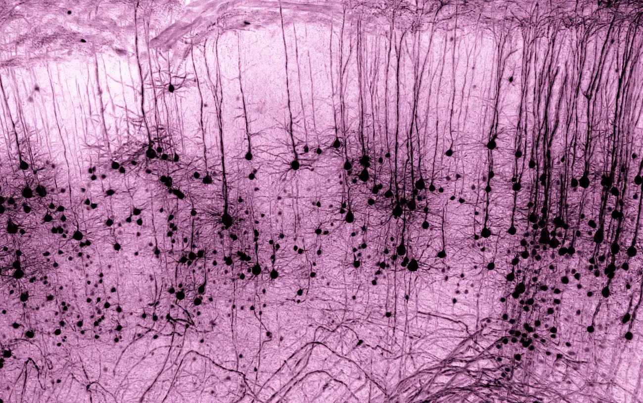
Images like the honorable mention of Bettina Rákóczi’s Alzheimer’s disease mouse model provide critical visual data for understanding neurodegenerative diseases. By using fluorescent immunostaining, researchers can track the progression of plaques in the brain tissue. Similarly, Marko Pende’s capture of a neuron in a tissue-cleared mouse brain demonstrates the power of "clearing" techniques, which make biological tissue transparent to allow for deeper 3D imaging. These advancements are vital for mapping the "connectome"—the intricate wiring of the nervous system.
Bridging the Communication Gap
One of the most significant implications of the Image of the Year Award is its ability to foster "Science Communication." In an era where scientific literacy is crucial, these images act as a hook. A person may be drawn to the "smiling" appearance of grass under a microscope or the "sand dunes" of lignin fiber, and through that curiosity, they learn about plant physiology or carbon sequestration.
Technological Inspiration
For the microscopy industry, these awards serve as a "stress test" for their equipment. When scientists like Igor Siwanowicz or Kentaro Mochizuki push their microscopes to capture these high-fidelity images, they provide feedback on the limits of current optical technology. This drives the development of next-generation lenses and sensors that will eventually lead to even more significant scientific discoveries.
Conclusion: A Window into the Infinite
The 6th Annual Evident Image of the Year Award reminds us that there is an infinite frontier right beneath our fingertips. From the jumping spider’s eyes captured by Walter Ferrari to the delicate fairyfly documented by Hanyang Xue, these images reveal a world of intense color, structural perfection, and biological drama.
As Katie Holden and her fellow winners receive their honors, the scientific community looks forward to the 7th year of the competition. With every passing year, the line between the scientist and the artist continues to blur, proving that to truly understand the world, one must be able to see—and appreciate—its inherent beauty.
For those interested in following the ongoing intersection of art and science, Evident Scientific maintains a gallery of past winners and honorable mentions on their official website and Instagram, providing a permanent archive of the world’s most beautiful microscopic discoveries.


Leave a Comment